
Figure 4.
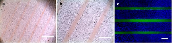
Optical photographs (a, b) and a fluorescence image (c) of fibroblasts cultured on a gelatin film that was photo-patterned with 8arm-PEG-CF(GPO)9. Cells exclusively attached to the unpatterned areas and formed a confluent monolayer. The immobilized CF-labeled PEG-CMP conjugate appears as clear orange (a, b) or green lines (c); the fibroblasts were fixed and stained with DAPI to visualize the nuclei (c). Scale bars: 1 mm in (a), 400 mm in (b), and 380 mm in (c).