Fig. 8.
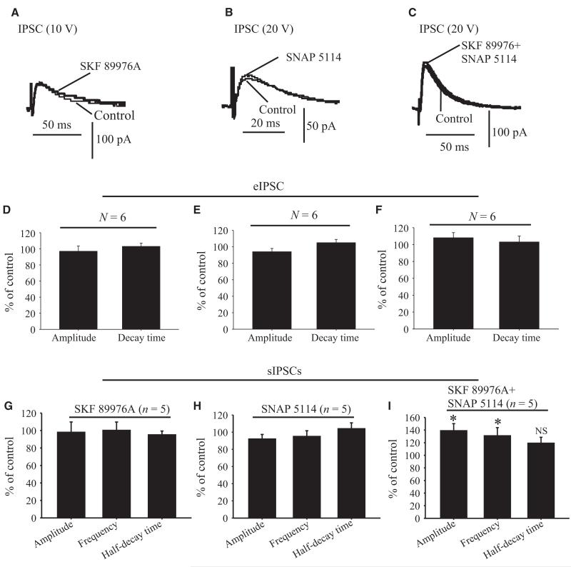
Effects of GAT-1, GAT-3 or GAT-1 combined with GAT-3 inhibitors on IPSCs evoked in GP neurons by local intrapallidal stimulation in coronal striatopallidal slice preparations. Application of SKF 89976A (A), SNAP 5114 (B) or SKF 89976A together with SNAP 5114 (C) has no significant effect on the amplitude and decay time of IPSCs evoked in GP neurons by local intrapallidal stimulation (20 V) in coronal slices of rat striatopallidal complex. (D–F) Summary bar graphs showing that the amplitude and decay time of eIPSCs in the rat GP are not changed in the presence of SKF 89976A (D), SNAP 5114 (E) or SKF 89976A together with SNAP 5114 (F) expressed as percentage of control ± SEM (P = 0.06). (G–I) Summary bar graphs showing that the amplitude, frequency and half-decay time of sIPSCs are not altered in presence of SKF 89976A (G) or SNAP 5114 (H) alone, but that their amplitude and frequency are significantly increased in the presence of SKF 89976A together with SNAP 5114, expressed as percentage of control ± SEM (*P = 0.001) (I). N, number of cells tested; NS, not significant.

