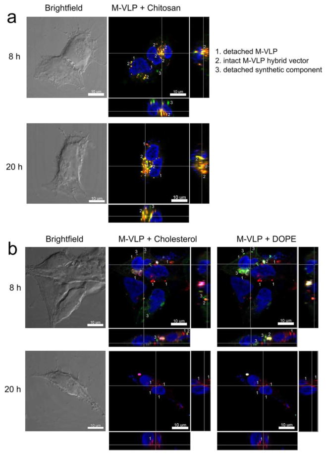
Figure 6.
Confocal fluorescence microscopy of HEK293 cells transfected with (a) Chi/M-VLP (5 μg/109 M-VLPs) or (b) Lip587/M-VLP (10 μg/109 M-VLPs) at 8 and 20 h post-transfection. Fluorescent labels were DiD (M-VLPs, red), RITC (chitosan, green), Rhod-B (DOPE, green) and NBD-6-cholesterol (green). Cell nucleus was counterstained with DAPI (blue). Scale bar = 10 μm.

