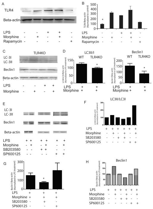
Figure 2. Morphine potentiates LPS-induced autophagy initiation through TLR4-p38 signaling.
(A) Morphine increased LPS-induced TLR4 expression in WT BMDMs. (B) Densitometric analysis of TLR4 expression. Values represent percentages of TLR4 normalized against beta-actin and compared with untreated control. *, P<0.05, Shown are representative results from one of three independent experiments. (C) LC3, Beclin1 and Bcl-2 expressions in BMDMs derived from TLR4KO mice following treatment with morphine in the presence or absence of LPS. (D) Densitometric analysis of LC3II/I and Beclin1 expression levels in BMDMs harvested from WT and TLR4KO mice. Values were normalized against beta-actin and represented as percentage of LPS treatment. (E) WT BMDMs were incubated with p38 mitogen-activated protein kinases(MAPK) inhibitor SB203580 (20μM) or c-Jun N-terminal kinases (JNK) inhibitor SP600125 (20μM) for 2 hours before treatment with morphine for 24 hours. Cells were then treated with LPS for 24 hours. LC3 and Beclin1 protein levels were evaluated using Western blot. (F and H) Densitometric analysis of LC3II/I and Beclin1 expressions in the representative blot. (G) Statistical analysis of Beclin1 levels from three independent blots in the presence or absence of MAP kinase inhibitors SB203580 (20μM) or c-Jun N-terminal kinases (JNK) inhibitor SP600125 (20μM). Values were normalized against beta-actin and represented as percentage of LPS treatment. The data suggested that p38 MAPK signaling plays a crucial role in morphine modulation of LPS-induced autophagy.

