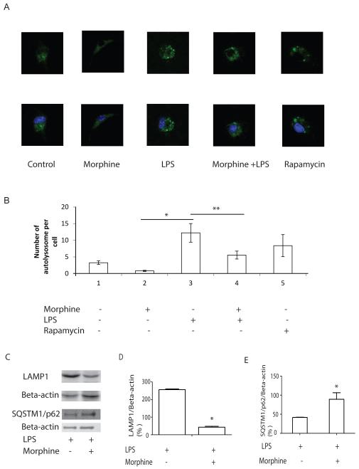
Figure 3. Morphine inhibits LPS-induced maturation of autophagosomes.
(A) Upper panel: BMDMs were transiently transfected with GFP-LAMP1 and cultured for 24 hours. Cells were then treated with morphine for 24 before LPS treatment for 24 hours. Images were visualized by fluorescence microscope at 600×magnification. Lower panel: Nuclei was labeled with DAPI. (B) Quantification of the number of autophagosomes in a single cell. GFP-LAMP1 vesicles in a single cell was counted. GFP-LAMP1-labeled cells from different treatment groups were merged with DAPI. *, P<0.05. **, P<0.05. (C) LAMP1 and SQSTM1/p62 protein levels were evaluated using Western blot. (D) Densitometric analysis of LAMP1 expression. Values were normalized against beta-actin. *, P<0.05, replicated experiments gave similar statistical significances. (E) Densitometric analysis of SQSTM1/p62 expression. Values were normalized against beta-actin. *, P<0.05, Representative data are from three independent experiments. Morphine inhibited phagolysomal maturation indicated by inhibition of LPS-induced LAMP1 expression and prevention of p62 degradation.

