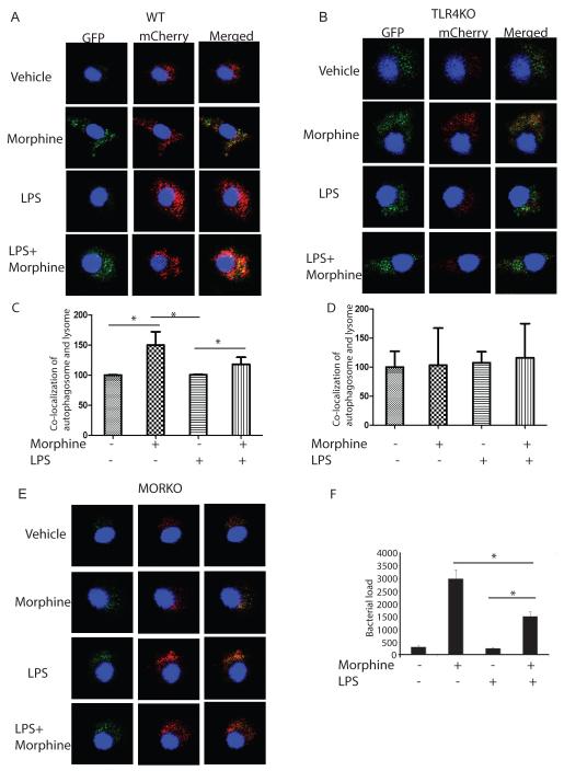
Figure 4. Morphine inhibits LPS induced autophagolysosome fusion.
(A, B and E) BMDMs from WT, TLR4KO and MORKO were transfected by GFP-mCherry-LC3, separately. Twenty-four hours after transfection, cells were treated with morphine for 24 hours before LPS treatment. Cells were then fixed before being subjected to confocal microscopy. (C) Quantification of co-localization of autophagosome and lysosome in WT mice BMDMs. *, P<0.05. (D) Quantification of co-localization of autophagosome and lysosome in TLR4KO mice BMDMs. (F) BMDMs were treated with morphine overnight and then treated with LPS for 24 hours. BMDMs were then infected with S. pneumoniae for 60 mins. Bacterial killing was allowed to occur for another 24 hours. Viable internalized bacteria within the cells were enumerated by a colony forming unit (CFU) assay. Results from three independent experiments are presented. *, P<0.05. In the presence of LPS, morphine treatment significantly increased S. pneumoniae load compared to LPS treatment.

