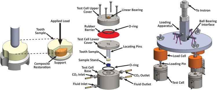
Fig 1.

(a) Schematic of the simulated tooth filling sample and the loading configuration; at right, the dentin is made transparent to observe the composite disk and support ring. (b) Exploded view of a bioreactor test cell. (c) Schematic of the load distribution system for loading three bioreactor test cells simultaneously.
