
Figure 4. Biodistribution of DIL-labeled exosomes in mouse brain.
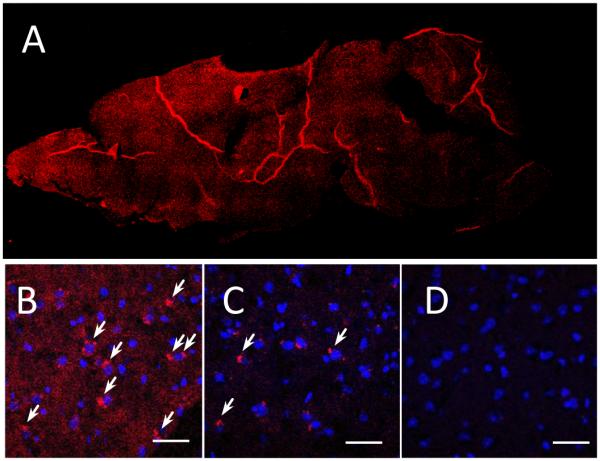
Exosomes were administered to mice with 6-OHDA-induced brain inflammation through: intranasal (A, B), or intravenous (C) routs; and compared to PBS-injected controls (D). The bar: 40 µm.