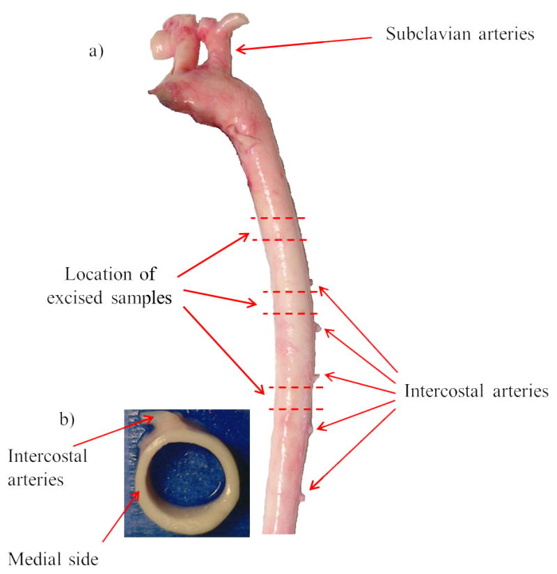
Figure 1.

a) Location of excised samples along the descending thoracic porcine aorta. b) Top view of a cylindrical section. Location of experiments across the wall thickness in the medial side of aorta is shown which is at 90° counter-clockwise with respect to intercostal arteries toward the heart.
