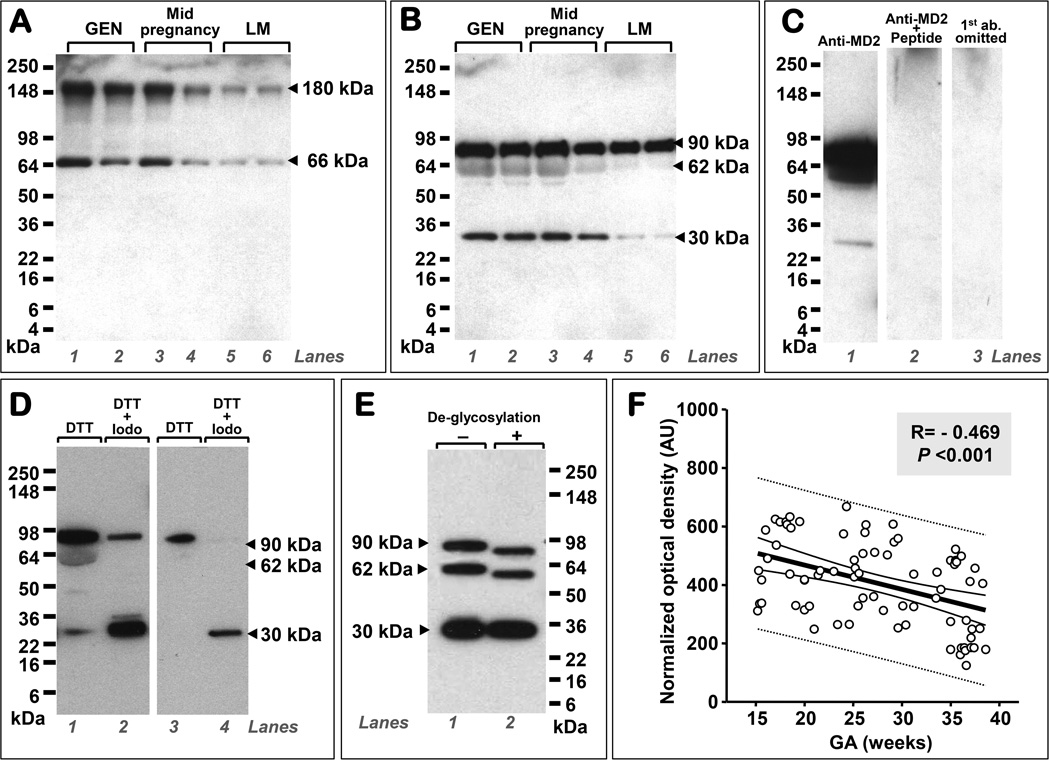
Figure 3. Human amniotic fluid (AF) contains soluble myeloid differentiation-2 (sMD-2) monomeric and multimeric proteoforms, is glycosylated, and its levels are gestational age (GA) regulated.
The figure is a composite of Western blot data demonstrating that the immunoreactivity of human AF sMD-2 in non-reducing conditions is characterized by two bands located at ~180 and ~66 kDa, respectively. The polyclonal rabbit anti-MD-2 primary Ab raised against a peptide corresponding to amino acids near the middle region of human MD-2 detected the two MD-2 forms in samples of AF retrieved by amniocentesis during the second trimester for genetic (GEN, n-26) purposes (A, lane 1 and 2), second trimester for symptoms of preterm labor in patients that delivered at term (n=50, A, lanes 3 and 4), and third trimester in patients who were tested for fetal lung maturity (LM, n=26) (A, lanes 5 and 6). Under reducing conditions, the same Ab detected three specific bands at 90, 62, and 30 kDa (B). Representative Western blot gels of AF retrieved during the second trimester for genetic (GEN) purposes (B, lanes 1 and 2), second trimester in patients with symptoms of preterm labor, negative microbial cultures who delivered at term (B, lanes 3 and 4), and third trimester for lung maturity (LM) testing (B, lanes 5 and 6). Each lane represents a sample from a different woman. Specificity of the sMD-2 bands was confirmed (C). Shown is a representative Western blot using the anti-MD-2 Ab in a second trimester AF genetic sample (C, lane 1), by pre-adsorbing the primary antibody with neutralizing peptide (C, lane 2) and by omitting the primary antibody (C, lane 3). Reduction+alkylation experiments performed using second trimester genetic amniocentesis fluid showed that the 90 and 62 kDa bands hold MD-2 multimers (D). As shown the molecular forms of sMD-2 migrating at 90 and 62 kDa (D, lane 1) diminished in intensity (D, lane 2) with the concurrent increase in intensity of the 30 kDa isoform (D, lane 2). This shift was more apparent if less AF protein was subjected to reduction+alkylation (D, lanes 3 and 4). Deglycosylation experiments produced a shift in the electrophoretic mobility of the 90 and 62 kDa bands (E, lane 1 vs. lane 2) but not of the 30 kDa band. Densitometric image analysis of the 90, 62, and 30 kDa polypeptides demonstrated that AF sMD-2 levels decrease with increasing gestational age (GA) (F). The thick black line represents the linear regression line, the thin black lines mark the confidence interval and the dotted lines show the prediction interval. Abbreviations: dithiothreitol (DTT), Iodo (iodoacetamine), AU: arbitrary densitometric units.

