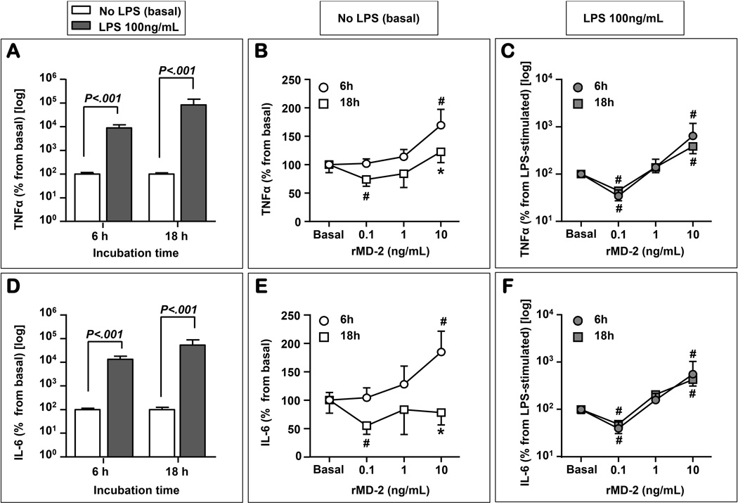
Figure 8. TNF-α and IL-6 release is stimulated by LPS and influenced by recombinant soluble myeloid differentiation-2 (rMD-2) treatment.
LPS (100 ng/mL) treatment significantly stimulated the production of TNF-α after 6h and 18h of stimulation (A). In a time and dose dependent manner, and in the absence of LPS, rMD-2 impacted on the release of TNF-α (B). At 6 hours, the 10 ng/mL dose of rMD-2 resulted in a significant increase in the TNF-α level. This effect was no longer seen at 18 hours. Conversely, the 0.1 ng/mL rMD-2 had an inhibitory effect 18h after treatment. In the presence of LPS, the inhibitory effect of the 0.1 ng/mL and the stimulatory effect of the 10 ng/mL rMD-2 dose were seen at both 6h and 18h, respectively (C). LPS treatment significantly stimulated the production of IL-6 after 6h and 18h of stimulation (D). At 6 h, in the absence of LPS, the 10 ng/mL dose of rMD-2 significantly increasing the IL-6 levels (E). In contrast, the 0.1 ng/mL rMD-2 had an inhibitory effect 18h after treatment. In the presence of LPS, at both 6h and 18 h, the 0.1 ng/mL of rMD-2 inhibit the release of IL-6 (F), while the 10 ng/mL dose displayed a stimulatory effect (F). * P<0.05 vs. 6h time point; # P<0.05 vs. basal (no rMD-2).

