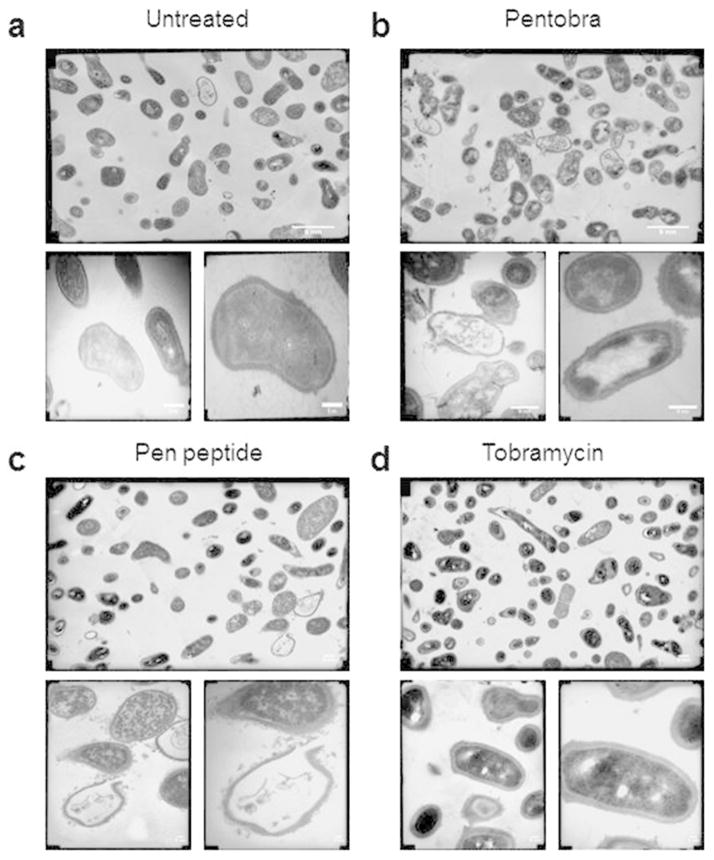
Figure 3. EM studies show Pentobra acts on P. acnes cell membranes.
Representative TEM micrographs of (A) untreated P. acnes control, (B) P. acnes after 3 h incubation with 25.7μM Pentobra, (C) P. acnes after 3 h incubation with 25.7μM pen peptide, and, (D) P. acnes after 3 h incubation with 25.7μM tobramycin. TEM were imaged at 10K magnification (top), 36K (bottom left), and 72K (bottom right). Compared with untreated control, the bacteria exposed to Pentobra and pen peptide exhibit cellular differences indicative of stresses on the membrane. Complete lysis of the cell membrane occurs, and the envelope boundary is now decorated with numerous blebbing events (bottom images in B and C). Scale bar = 5nm.

