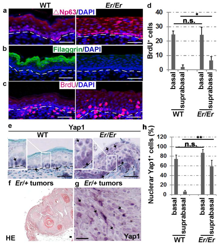
Figure 1. Enriched nuclear localization of Yap1 in Er/Er epidermis and induced skin tumors.
(a–c) Immunofluorescence staining for ΔNp63 (a), filaggrin (b) and BrdU (c) in the E18.5 Er/Er epidermis (right) compared with the WT control (left). (d) Quantification of BrdU+ cells. The number of BrdU+ cells per 500 μm of fixed length parallel to the epidermis surface was counted. Two-tail t-test: *p<0.05, N=5 embryos. n.s., not significant. (e) Immunohistochemical staining of Yap1. H&E staining (f) and Yap1 cellular localization (brown) detected by immunohistochemical staining (g) of DMBA/TPA-induced tumors derived from Er/+ mice. The scale bars represent 50 μm in (a–f) and 150 μm in (g). The arrows indicate Yap1-positive cells in (e) and (g). (h) Quantification of nuclear Yap1-positive cells. Two-tail t-test: **p<0.01, N=3 embryos.

