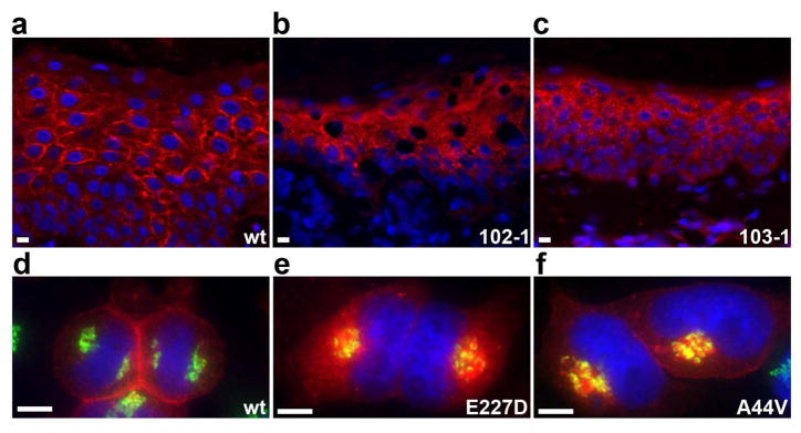
Figure 6. Cx43 is mis-localized in EKVP.

(a–c) Cx43 immunolocalization was performed on tissue sections from wild-type (wt) and affected patient skin (102-1: E227D, 103-1: A44V), with Cx43 antibody in red and DAPI nuclear counterstain in blue. (a) Wild-type tissue shows primarily intercellular membrane localization of Cx43. (b–c) Affected skin shows primarily cytoplasmic localization. (d–f) Wild-type (wt) and mutant Cx43 (E227D, A44V) were HA-tagged and expressed in HeLa cells. Immunolocalization of Cx43 is red, cis-Golgi marker GM130 is green, and DAPI nuclear counterstain is blue. (d) Wild-type Cx43 localizes to intercellular junctions and does not co-localize with GM130. (e and f) The E227D and A44V mutants do not localize to intercellular junctions and accumulate in a subcellular compartment, partly co-localizing with GM130. Scale bars are 20 μm in all panels.
