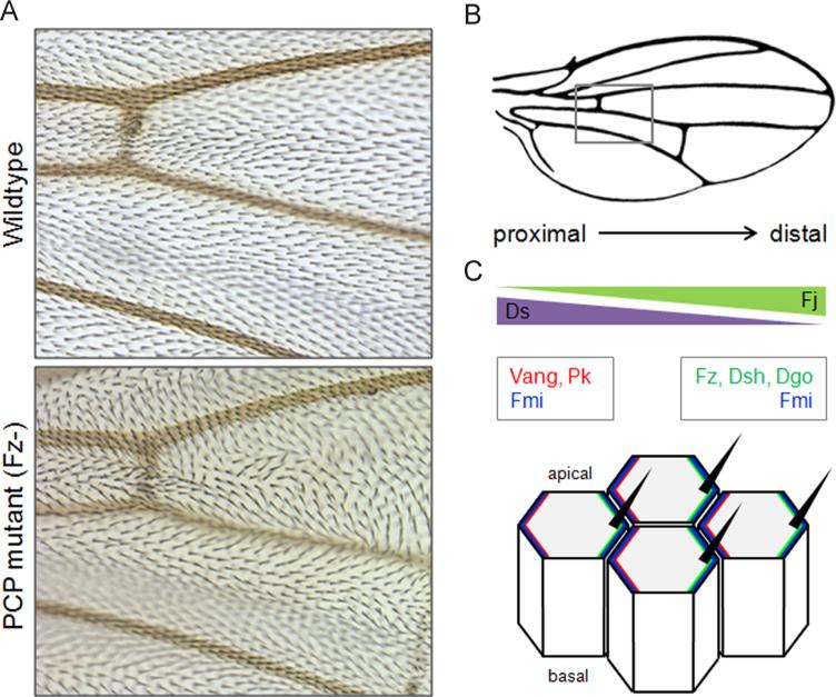
Fig. 1.
PCP signaling in the Drosophila wing. A) Epithelial cells in the wing blade generate an actin hair pointing distally in wildtype flies, while mutants lacking Fz show disturbed hair polarization with swirls and waves. The original images were kindly provided by Marek Mlodzik and Jun Wu. B) Schematic illustration of a wing. The grey box indicates the region shown in A. C) Global PCP components Ds and Fj are expressed in opposing gradients across the wing, while the core PCP proteins are asymmetrically localized at the cell junctions between neighboring cells. The asymmetries of global and core proteins generate tissue polarity.

