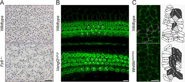
Fig. 2.
Examples of vertebrate PCP. A) Hair and hair follicles in dorsal skin of wildtype and Fz6−/− mutant mice at P3, visualized with melanin pigmentation. Mice are oriented with anterior to the left and posterior to the right. In PCP mutants, hairs do not point distally as in but lose their uniform polarity. Scale: 0.5 mm. Images were kindly provided by Jeremy Nathans and Hao Chang. B) Orientation of sensory hair cells of the cochlea (inner ear) of E18.5 wildtype and Vangl2 mutant mice. Polarized bundles of stereocilia are uniformly oriented in wildtype mice, while their orientation becomes randomized in the PCP mutant (direction indicated by white arrows). Scale: 10 μm. Original images were kindly provided by Matthew Kelley. C) Polarized orientation of tubule cells perpendicular to the axis of extension is disturbed in Wnt9b mutants. Confocal images (single focal plane from Z-stack) show collecting ducts in E15.5 wildtype and Wnt9bneo/neo kidneys, immunostained for E-cadherin (green), DBA (collecting duct marker; magenta) and Par3 (apical membrane marker; red). Chosen areas represent regions just basal to the apical membrane, identified by absence of Par3. Cell outlines: white cells are perpendicular to the axis of elongation (45–90%), dark gray cells are parallel (0–45%). Scale: 10 μm. See Karner et al. [54] for quantification.

