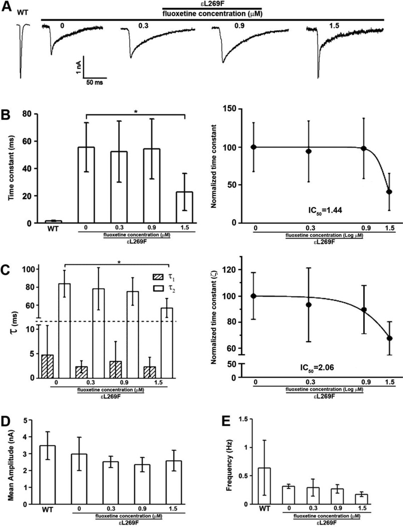
Figure 1. Fluoxetine treatment corrects altered mSCS εL269F nAChR kinetics.
Two-electrode voltage clamp (TEVC) recordings of quantal activity for from wild type and εL269F mice diaphragms treated acutely with fluoxetine at concentrations of 0.3, 0.9 and 1.5 µM. A. The decay time constant in control SCS transgenic mice was 55.60 ± 17.95 ms whereas 52.48 ± 22.26 in 0.3 µM treatment, 54.56 ± 22.04 in 0.9 µM treatment and 22.80 ± 13.59 in 1.5 µM treatment. B. The MEPC time constant was dramatically reduced in SCS transgenic mice with 1.5 µM treatment. C. Decay phases were resolved into two exponents (τ1 and τ2) with τ2 showing a concentration dependent reduction. n=4, *p<0.05. Student’s t-test. D and E. Neither mean amplitude nor the frequency of MEPCs recorded from εL269F mice was affected by fluoxetine. n=4; Student’s t-test.

