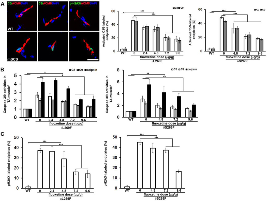
Figure 5. Fluoxetine prevents pathological activation of calpain, caspase-3 and caspase-9 at NMJs, and subsynaptic DNA damage in mSCS.
A. Left. Co-localization of activated caspase-3 (green, arrows) and caspase-9 (green, arrows) with NMJs (red) in WT and mSCS. Activated caspase-3 and caspase-9 are seen in mSCS NMJ with subsynaptic nuclei, but are not seen in WT. Ca2+-mediated DNA damage in subsynaptic nuclei was exhibited as co-localization of phospho-H2AX-labeled (pH2AX, green) nuclei (DAPI, blue) with NMJ (red) in untreated mSCS. n=5; Scale bar=15µm. Middle. Quantitation of endplates labeled with activated caspase-3 and caspase-9 in WT, untreated mSCS (εL269F) and mSCS (εL269F) treated with fluoxetine under indicated conditions. In untreated mSCS (εL269F) 45.9±5.0% of endplates were labeled with cleaved caspase-3 and 45.2±5.9% with cleaved caspase-9, while no active caspases were detected in WT endplates. Treatment of mSCS (εL269F) muscle with 9.6µg/g fluoxetine significantly reduced labeling for cleaved caspase-3 (18.2±4.1%) and caspase-9 (16.1±4.1%). Right. Compared with cleaved caspase-3 (48.3±1.8%) and cleaved caspase-9 (42.8±2.0%) in untreated mSCS (δS268F), fluoxetine treatment with 9.6µg/g significantly reduced labeling for cleaved caspase-3 and caspase-9 to 18.3±1.5% and 16.7±2.1%, respectively. n=5; ***p<0.001. All comparisons were analyzed by Student’s t-test.
B. Relative activity of calpain, caspase-3 and caspase-9 proteases (# = normalized to WT) for untreated mSCS and treated mSCS muscle under indicated conditions. Left. Protease activities for untreated mSCS (εL269F) muscle are 4.2-fold (calpain), 2.7-fold (caspase-3), and 2.0-fold (caspase-9) to WT activity while reduced protease activities of 2.2-fold (calpain), 1.3-fold (caspase-3) and 1.3-fold (caspase-9) in εL269F treated with 9.6µg/g fluoxetine. n=5; *p<0.05. Right. Protease activities for untreated mSCS (δS268F) muscle are 5.5-fold (calpain), 3.1-fold (caspase-3), and 2.4-fold (caspase-9) to WT activity while reduced protease activities of 2.1-fold (calpain), 1.5-fold (caspase-3) and 1.2-fold (caspase-9) in mSCS (δS268F) with 9.6µg/g fluoxetine treatment. n=7; **p<0.01. All comparisons were analyzed by Mann-Whitney u-test.
C. Quantitation of pH2AX-labeled NMJs in WT, untreated mSCS and mSCS treated with fluoxetine under indicated conditions. Left. In WT NMJ pH2AX-labeled nuclei were rare (1.4±1.2%). In untreated mSCS (εL269F) a significant number of NMJ nuclei were labeled with pH2AX (37.3±2.2%). Treatment of mSCS (εL269F) with 9.6µg/g fluoxetine significantly reduced pH2AX-labeling to 14.3±3.7%. Right. In untreated mSCS (δS268F) a significant number of NMJ nuclei were labeled with pH2AX (45.1±2.3%). Treatment of δS268F with 9.6µg/g fluoxetine significantly reduced pH2AX-labeling to 16.6±1.5%. εL269F=5; δS268F=7; ***p<0.001. All comparisons were analyzed by Student’s t-test.

