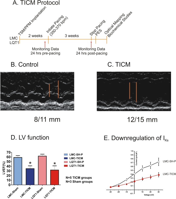
Fig 1. TICM Protocol.
(A) Three-week pacing protocol. TSM/PPM = transmitter/ pacemaker, PES = programmed electrical stimulation. (B) TICM groups show dilated left ventricle and reduced ejection fraction. The red bars indicate end-systolic and end-diastolic LV internal dimensions (8 and 11 mm for LMC sham pacing and 12 and 15 mm for TICM). (C) Post-pacing protocol LV function presented as left ventricular ejection fraction in LMC-TICM, LQT1-TICM, and their sham controls. TICM rabbits show statistically significant differences in LV ejection fraction compared with sham; *P<0.05. (D) Quantification of IKs in LMC rabbits. Current amplitude was normalized to cell capacitance. Compared with LMC-SH-P (n = 12), significant downregulation of IKs was seen in LMC-TICM (n = 11).

