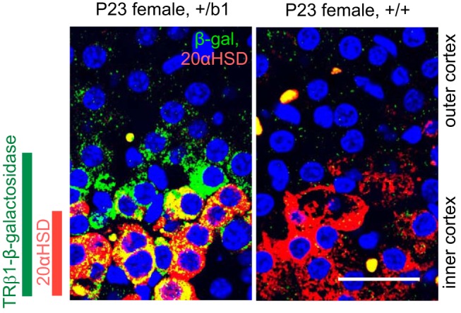
Figure 2.

TRβ1-positive zone incorporates 20αHSD-positive zone in the adrenal inner cortex. Confocal image of double-immunofluorescence analysis for β-galactosidase (green) and 20αHSD (red) in adrenal sections of female mice. In +/b1 mice, the TRβ1-positive (β-galactosidase) cell zone is broader than and incorporates the 20αHSD-positive cell zone (orange or yellow, doubly positive cells). Specific β-galactosidase signal was not detected in +/+ control mice. DAPI (blue) marks all cell nuclei. Scale bars, 25 μm.
