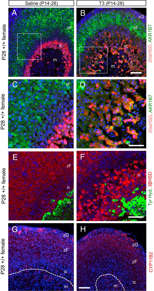
Figure 4.

T3-induced changes in cortical marker expression in the inner cortex. A and B, Immunofluorescence analysis showing that T3 treatment expanded the 20αHSD-positive cell zone (red) and in the innermost cortical zone induced coexpression of the outer cortical, zona fasciculata marker AKR1B7 (green, or yellow for double positive). Mice were treated with T3 or saline from P14 to P28. C and D, Representative higher magnification of panels A and B showing cells positive for both 20αHSD and AKR1B7. E and F, Immunofluorescence analysis showing prominent expression of steroidogenic enzyme 3βHSD (red) in the expanded inner zone next to the medulla (green, tyrosine hydroxylase) after T3 treatment. G and H, Immunofluorescence analysis showing normal expression of CYP11B2 in the zona glomerulosa under saline or T3 treatment. In all panels, DAPI (blue) marks cell nuclei. Abbreviations are the same as in Figure 3. Scale bars for panels A, B, G, and H, 100 μm; for C–F, 50 μm.
