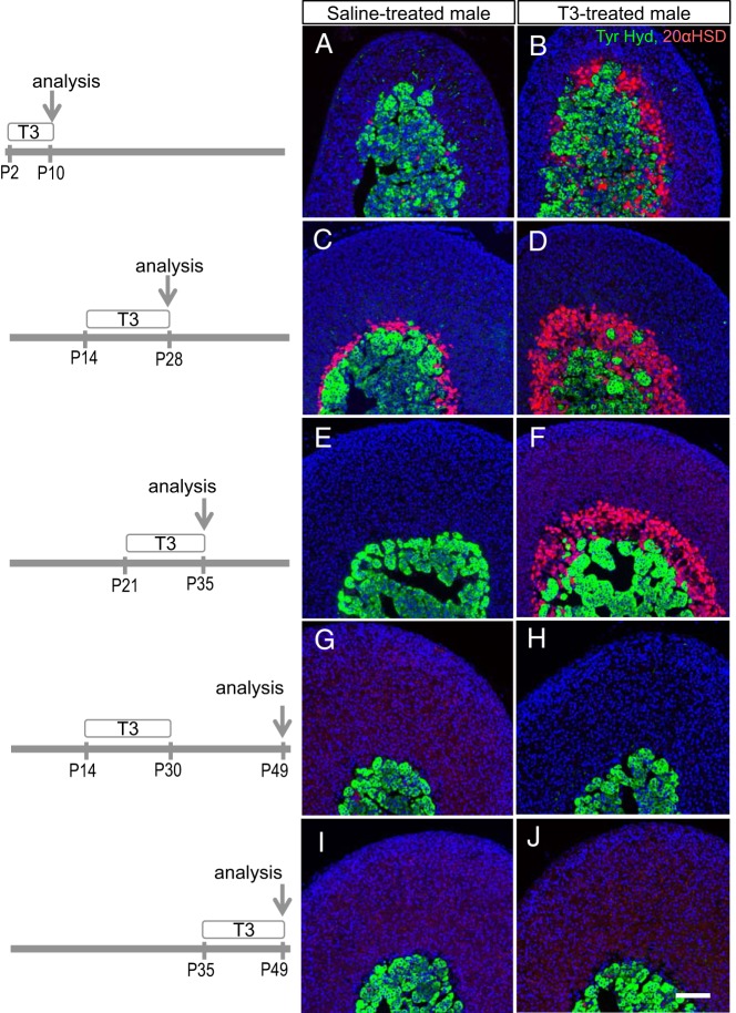
Figure 5.
T3 influence on the appearance and regression of the 20αHSD-positive inner cortex. Periods of treatment of male mice with T3 or saline (postnatal days) and day of analysis are shown on the left. A and B, T3 resulted in premature appearance of a 20αHSD-positive zone (red) at P10. C and D, T3 stimulated expansion of the 20αHSD-positive zone at P28. E and F, T3 prolonged the presence of the 20αHSD-positive zone at P35. G and H, Removal of T3 treatment after P30 resulted in loss of 20αHSD-positive zone. I and J, T3 given after regression of the 20αHSD-positive zone did not recover a 20αHSD-positive zone. Tyr. Hyd., tyrosine hydroxylase (medulla marker, green). DAPI (blue) marks cell nuclei. Scale bars, 100 μm.

