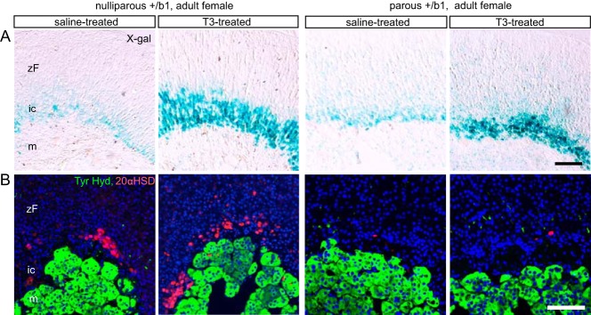
Figure 6.

T3 sensitivity of the TRβ1-positive inner cortex in nulliparous or parous adult female mice. A, X-gal staining showing expansion of the TRβ1-positive inner cortex in response to T3 in both nulliparous and parous adult female mice (6 mo old, given T3 or saline for 2 wk before analysis). B, Immunofluorescence analysis showing substantial loss of 20αHSD expression after pregnancy (parous) and the inability of T3 to recover 20αHSD in parous mice. DAPI (blue) marks cell nuclei. Scale bars, 100 μm. ic, inner cortex; m, medulla; zF, zona fasciculata.
