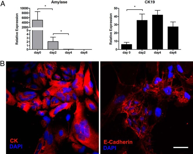
Figure 1.
Characterization of isolated exocrine cells. A, Changes in the gene expression profile of exocrine cells 0, 2, 4, and 6 days after isolation. Freshly isolated exocrine cells (d 0) had high expression of amylase, which disappeared in just 4 days. The results were obtained from the adherent cells after floating cells were removed on each day except day 0 (freshly isolated nonadherent exocrine cells). Mean ± SEM, 4 independent experiments (each with duplicates). *, P < .05. B, Seven days after isolation, the adherent cells had proliferated and formed epithelial-like monolayers with cobblestone-like morphology; immunostaining was for pan-CK (red) (left panel) and E-cadherin (red) (right panel). Blue represents nuclear staining with 4′,6-diamidino-2-phenylindole (DAPI). Scale bar, 50 μm. Images are representative of 4 independent experiments.

