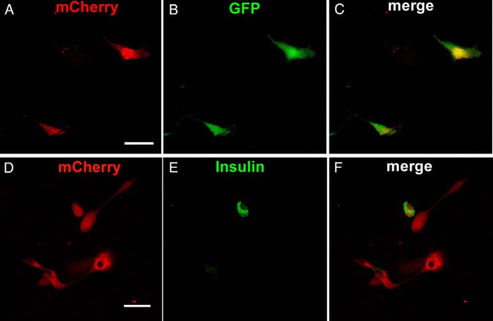
Figure 3.
GFP and insulin expression in Ad-M3C-transduced cells 7 days after transduction. A–C, Coexpression of mCherry and GFP in cells from MIP-GFP mice fixed 7 days after Ad-M3C transduction and examined with confocal microscopy. Some mCherry+ (red)-transduced cells also expressed GFP fluorescence (green), indicating activity of the insulin promoter. Merged image (yellow). (D-F) Ad-M3C-treated mCherry+ (red) cells from DBA/2 mice were immunostained for insulin (green) 7 days after transduction. Merged images (yellow) showed coexpression of mCherry and insulin in some but not all Ad-M3C-transduced cells. Images are representative of 3 independent experiments. Scale bars, 50 μm.

