Figure 1.
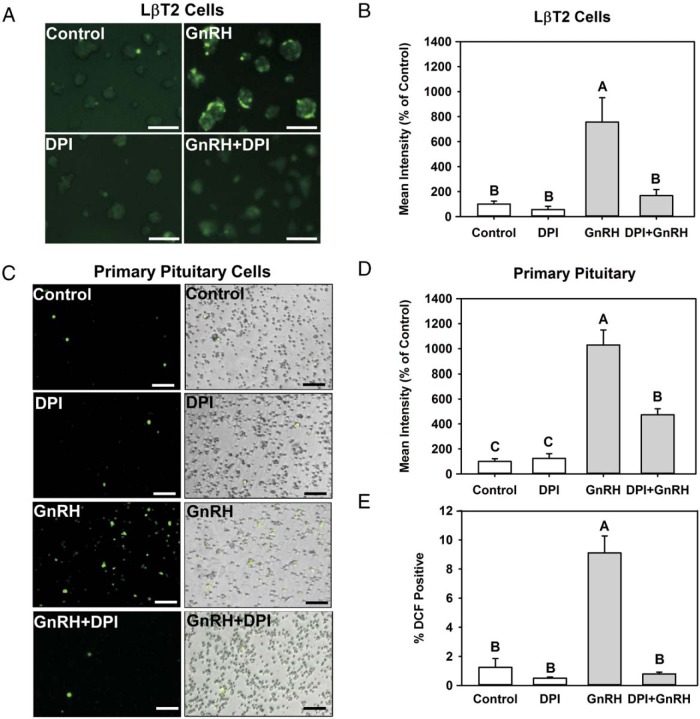
GnRH stimulation induces intracellular ROS production via NOX/DUOX activity. A–D, Serum-starved LβT2 cells (A) and female mouse primary pituitary cells (C) were preincubated with vehicle (dimethylsulfoxide) or 5 μM DPI for 30 minutes. Afterwards, cells were exposed to either vehicle (control) or 10 nM GnRH for 30 minutes and then treated with 10 μM CM-H2-DCFDA for 30 minutes. Intracellular ROS generation was visualized by fluorescence microscopy (A and C) and quantified by image analysis (B and D). E, Percentages of cells responsive to GnRH stimulation were quantified and reported relative to total cell numbers. In LβT2 cells, GnRH alone stimulated levels 756% over control, whereas DPI-treated cells responded only 168%. DPI alone fluoresced 84% of control, but this was not significantly different. In primary pituitary cells, DPI significantly inhibited GnRH stimulation 1024% over control with GnRH vs 474% over control in DPI+GnRH-treated cells. Results represent means ± SEM from 3 independent experiments normalized with the control. Scale bars correspond to 150 μm. Letters represent significant differences (P < .05) by ANOVA followed by the Tukey multiple comparison test. Groups with different letters are significantly different from each other.

