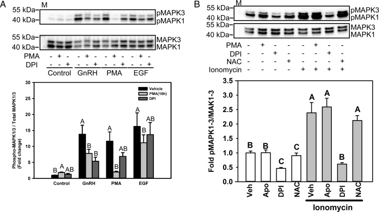
Figure 4.
GnRH-mediated MAPK1/3 activation is regulated by the PKC/NOX pathway. A, Serum-starved LβT2 cells were preincubated with 1 μM PMA for 16 hours or with 5 μM DPI for 30 minutes. After treatment, the cells were incubated with 10 nM GnRH, 100 nM PMA, or 50 ng/mL EGF for 5 minutes. The cells were lysed and analyzed by Western blotting for phosphorylated (p) MAPK1/3. The blots were stripped and reblotted for total MAPK1/3. The graph shows the quantitative chemiluminescence results from 4 independent experiments. The data represent mean values ± SEM normalized to the vehicle control of 4 independent determinations. Data from vehicle or 5-minute GnRH, PMA, or EGF treatment groups were analyzed by ANOVA and post hoc testing with the Tukey multiple comparison test. Groups with different letters are significantly different from each other (P < .05) within treatment groups. B, Serum-starved LβT2 cells were preincubated with 500 μM apocynin, 5 μM DPI, or 10 mM NAC for 30 minutes. Cells were then treated with 1 μM ionomycin for 5 minutes. The cells were lysed and analyzed by Western blotting for phosphorylated MAPK1/3. The blots were stripped and reblotted for total MAPK1/3. The graph shows quantitative chemiluminescence results from 3 independent experiments. The data represent mean values ± SEM normalized to the vehicle control from 4 independent determinations. Groups with different letters are significantly different from each other (P < .05) by ANOVA followed by the Tukey multiple comparison test. M, Protein size marker.

