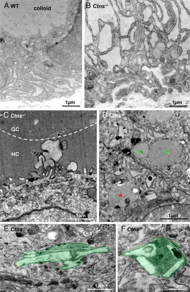
Figure 4.
Ultrastructural alterations in Ctns−/− thyrocytes. Representative EM views of resting WT (A) and activated Ctns−/− thyrocytes (B–F). A, In this resting WT thyrocyte at 12 months, notice limited ER expansion and thin apical projections. B, Hyperplastic Ctns−/− thyrocyte at 12 months showing strong dilation of endoplasmic reticulum lumen. Notice characteristic tortuous basal plasma membrane and basal lamina. C, This activated Ctns−/− thyrocyte at 8 months projects a lamellipodium, characteristic of macropinocytosis (arrowheads) across a homogenous peripheral colloid ring (HC) up to central granular colloid (GC), indicated by the broken line. D, Asterisks indicate colloid droplets in two adjacent activated thyrocytes, two with homogenous colloid content and undergoing homotypic fusion (green asterisks), one bearing additional luminal structures indicating fusion with lysosomes (red asterisk). E and F, Cystine crystals-bearing lysosomes in Ctns−/− thyrocytes at 12 months.

