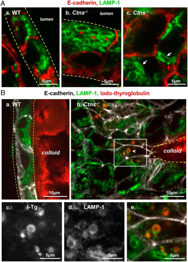
Figure 7.
Alterations of the late endocytic apparatus in hyperplastic Ctns−/− thyrocytes. A, Identification of large apical vacuoles and crystal-bearing structures as lysosomes. Comparison of WT (a) and Ctns−/− mice (b and c) at 9 months for immunolabeling of E-cadherin (red) and LAMP-1 (green; late endosome/lysosome membrane). a, In this resting WT follicle, flat thyrocytes show at their apical pole packed lysosomes of uniform small size and round shape. b and c, In activated Ctns−/− thyrocytes, late endosomes-lysosomes are frequently dilated (red arrowheads) and distorted (better seen at panel c, white arrows). Red arrows at panel c suggest docked but not fused late endosomes/lysosomes. For levels of Rab5 and Rab7 mRNAs, see Supplemental Figure 4. B, Iodo-Tg is retained in lysosomes. Comparison of WT (a) and Ctns−/− mice (b–e) at 9–10 months for E-cadherin (white), LAMP-1 (green), and iodo-Tg (red). In resting WT follicles (a), iodo-Tg is stored in the colloid and is never detected within thyrocytes. In activated Ctns−/− thyrocytes (b), vesicles filled with iodo-Tg and labeled by LAMP-1 (thus not primary colloid droplets) are frequently found at the apical pole of hyperplasic thyrocytes. Enlargements of the boxed field (c–e) first show single-channel images of iodo-Tg and LAMP-1 in black and white for optimal resolution and easier pattern comparison and then merged back with E-cadherin in triple colors as above. The apparent defect of (iodo-)Tg labeling of the central lumen is due to artifactual sticking of the colloid to the coverslip, thus out of focus by confocal imaging. For a gallery of representative images of iodo-Tg retained in lysosomes, see Supplemental Figure 3.

