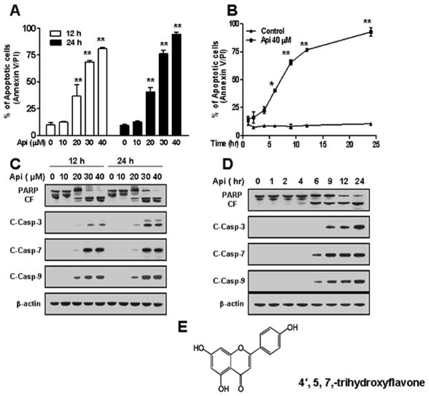
Fig. 1.
Effects of apigenin on apoptosis, caspases activation, and PARP cleavage in U937 cells. (A) U937 cells were treated without/with various concentrations of apigenin for 12 h and 24 h. (B) U937 cells were treated without/with 40 μM apigenin for indicated times. Cells were stained with Annexin V/ PI, and apoptosis was determined using flow cytometry as described in Materials and methods. The values obtained from Annexin V assays represent the means ± SD for three separate experiments. * or ** Values for cells exposed to apigenin were significantly increased compared to values in control cells by Student’s t-test; p < 0.05 or p < 0.01. (C–D) U937 cells were exposed to apigenin in dose and time-dependent manners. Total cellular extracts were subjected to Western blotting using indicated antibodies. (E) Structure of apigenin (4′, 5, 7,-trihydroxyflavone).

