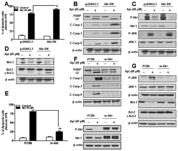
Fig. 4.
Determination of functional significance of Akt in apigenin-induced apoptosis. (A) U937 cells were transfected with an empty vector (pcDNA3.1) and Akt-DN as described in Materials and methods. Akt-DN expressing cells were treated without/with 40 μM of apigenin for 24 h, after which apoptosis was analyzed using Annexin V/PI assay. *Values for Akt-DN cells treated with apigenin were significantly increased compared to those for pcDNA3.1 cells by Student’s t-test; p < 0.05. (B-D) Total cellular extracts were subjected to Western blotting using indicated antibodies. (E) U937 cells were transfected with an empty vector (PCB6) and constitutively active myristolated Akt (m-Akt). m-Akt and PCB6 expressing cells were treated without/with 40 μM of apigenin for 24 h, after which apoptosis and P-Akt, and Akt1 expressions were analyzed. **Values for m-Akt cells treated with apigenin were significantly decreased compared to those for PCB6 cells by Student’s t-test; p < 0.01. (F-G) Total cellular extracts were subjected to Western blotting using indicated antibodies.

