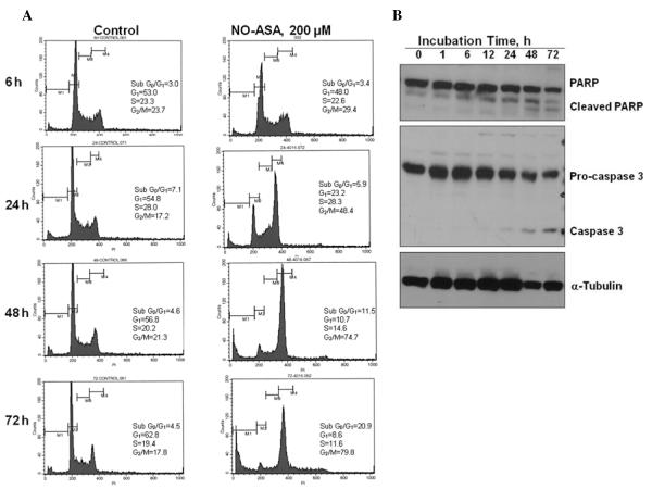
Figure 5.
NO-ASA-induced apoptosis in SW480 cells. (A), After SW480 cells were treated with either 200 μM NO-ASA or vehicle for the indicated periods of time, they were stained with PI and analyzed by flow cytometry. (B), SW480 cells were treated with 200 μM NO-ASA for the indicated periods of time, when proteins were extracted and analyzed by immunoblotting. Loading control: α-tubulin.

