
Figure 4.
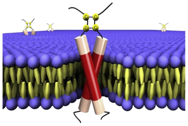
BPV1 E5 protein. A schematic representation of BPV1 E5 dimers embedded in a lipid bilayer. The transmembrane domain of the E5 protein is shown in red, with the disulfide-bonded carboxy terminus at the top (in the extracellular/luminal space). The membrane phospholipids are represented as spheres and the fatty acids as spindles. Reprinted from Reference 140a, copyright 2010, with permission from Elsevier.