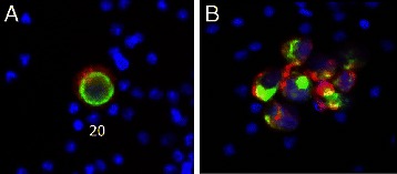
Figure 3.

Fluorescently stained model circulating tumor cells collected and imaged using the AccuCyte® – CyteFinder® system. (A) A549 mCTC stained with antibody to EGFR (red), cytokeratin (green), and nuclear dye (blue). (B) Cluster of LnCAP mCTCs stained with antibody to EpCAM (red), cytokeratin (green), and nuclear dye (blue). Cells imaged at scanning 10X objective magnification.
