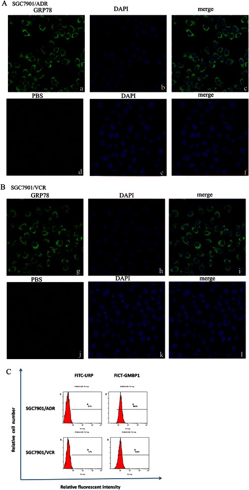
Figure 1.

Subcellular localization of GMBP1 and its receptor GRP78 in SGC7901/ADR and SGC7901/VCR. (A-B): a, d, g, j: The cytoplasmic localization of internalized GRP78 (green). b, e, h, k: Nuclear staining with 4, 6-diamidino-2-phenylindole (DAPI; blue). c, f, i, l: Merged images showing the relationship between GRP78 and the nucleus. (C): The internalization of the GMBP1 peptide into SGC7901/ADR and SGC7901/VCR cells. FITC-GMBP1 bound to SGC7901/ADR and SGC7901/VCR cells exhibited higher fluorescence intensity than the negative control FITC-URP group.
