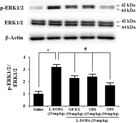
Figure 6.

Effects of GPS and GP-EX on L-DOPA-induced phosphorylation of ERK1/2 in 6-OHDA-lesioned rats. ERK1/2 phosphorylation (p-ERK1/2) was evaluated by western blotting of proteins extracted from the 6-OHDA-lesioned striatum 1 h after the final L-DOPA treatment. Immunoblot images were detected by antibodies against phospho-ERK1/2, ERK1/2 and β-actin using western blotting analysis. Values of the relative density ratios of p-ERK1/2/ERK1/2 are normalized and expressed in arbitrary units as compared with saline-treated group or L-DOPA alone-treated group. The position of molecular size markers is indicated as kDa. The results are expressed as mean ± S.E.M. for 8–10 animals/group. *P < 0.05 compared with saline-treated group; # P < 0.05 compared with L-DOPA alone-treated group (one-way ANOVA followed by Tukey’s test).
