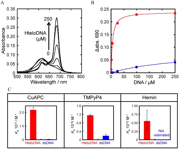
Figure 5.
(A) Visible absorbance spectra of 2.5 µM CuAPC with 0–250 µM of HteloDNA at 25 °C. The sample was first heated at 80 °C for 2 min, then gently cooled at 2 °C·min−1; (B) Absorbance of 2.5 µM CuAPC at 690 nm with 0-250 µM of HteloDNA and dsDNA (5'-AGAAGAGAAAGA-3'/5'-TCTTTCTCTTCT-3'); (C) Ka value of each ligand for HteloDNA and dsDNA. All measurements were carried out in a buffer containing 50 mM MES-LiOH (pH 7.0), 100 mM KCl, and 10 mM MgCl2 at 25 °C.

