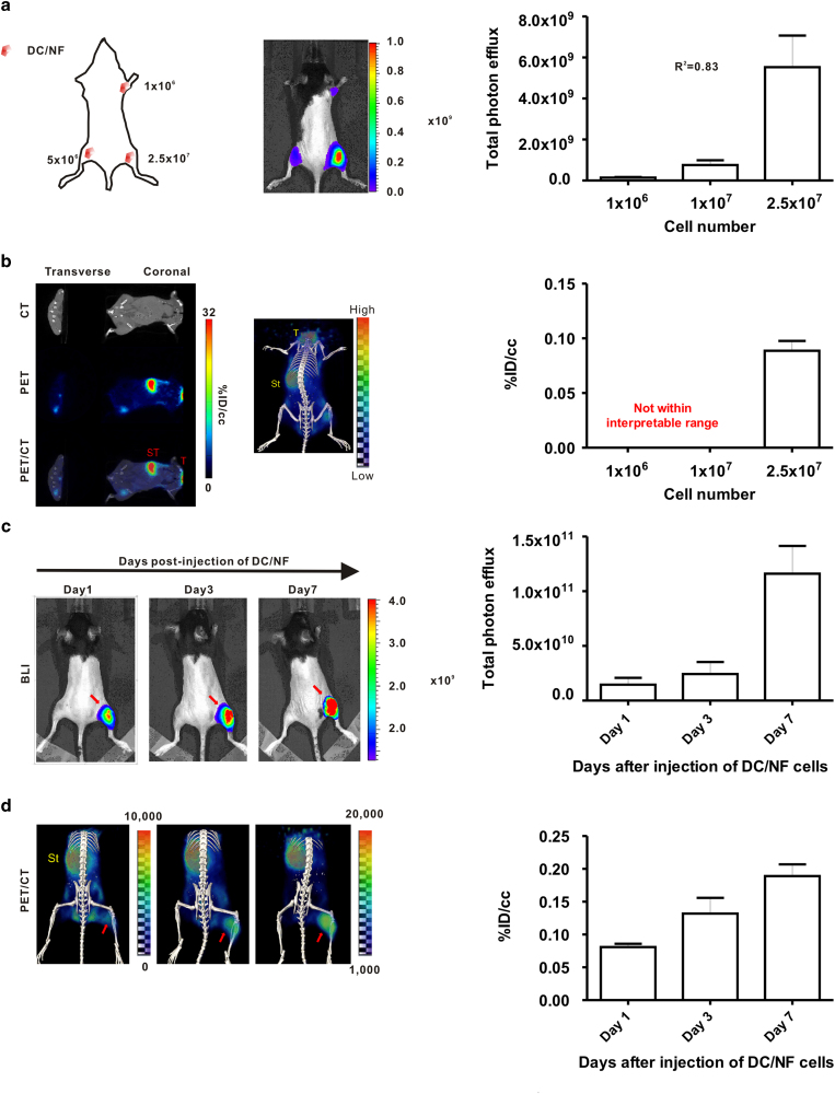
Figure 2. In vivo I-124 PET/CT imaging and BLI of DC2.4/NF cells in mice.
DC/NF cells were intramuscularly administered in the right upper thigh (1 × 10⁶), left lower thigh (5 × 10⁶), and right thigh (2.5 × 10⁷) of mice, and imaging was acquired. (a) In vivo BLI of DC/NF cells in mice. (b) In vivo I-124 PET/CT imaging of DC/NF cells in living mice. Mice received DC/NF cells by intramuscular injection into the right thigh, and imaging was acquired at the indicated times. In vivo visualization of the proliferation of infused DC/NF cells with both (c) BLI and (d) I-124 PET/CT in living mice. Physiological iodide uptake was observed in the thyroid (T) and stomach (ST). Red arrows indicate the site injected with DC/NF cells. The uptake of radioiodine in the region of interest was evaluated with PMOD software and is expressed as %ID/cc (percent injected dose per cc). Data are expressed as the mean ± standard deviation (SD) of 3 independent experiments (n = 5 mice).

