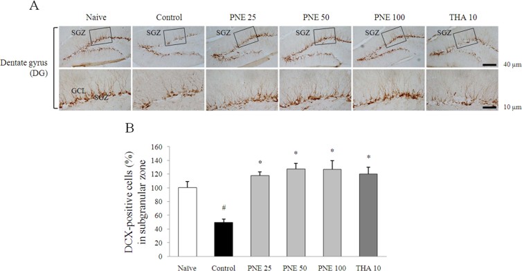
Figure 5. DCX immunohistochemical analysis of the effects of PNE on improved scopolamine-induced suppression of neurogenesis in the dentate gyrus.
(A) DCX-positive staining in immature neurons is shown in the subgranular zone of dentate gyrus. Representative photomicrographs were taken at magnifications of 100 and 400×. (B) Quantification of DCX population. Data are expressed as means ± SD (n = 3). #P < 0.05 compared with the naïve group; *P < 0.05, **P < 0.01 compared with the control group.

