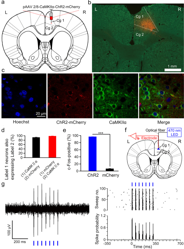
Figure 1. Selective labelling and optogenetic activation of the right caudal mPFC neurons.

(a) The rats were stereotactically injected with pAAV 2/8-CaMKIIα-ChR2-mCherry targeting the right caudal mPFC. (b) Example of ChR2-mCherry expression in the right caudal mPFC. (c) Representative images showing cell-specific ChR2-mCherry expression (red) in pyramidal neurons (green) of the right caudal mPFC. (d) Statistics of expression in the right caudal mPFC pyramidal neurons (502 cells, from five mice). (e) Percentage of c-Fos-positive cells among ChR2-mCherry-expressing cells (324/334 cells) or mCherry-expressing cells (21/320 cells) after light stimulation (n = 3 rats each; ***P < 0.001; two-tailed unpaired Student’s t-test). (f) In vivo right caudal mPFC “optrode” recording setup. (g) Multi-unit activity in the right caudal mPFC from a rat injected with pAAV 2/8-CaMKIIα-ChR2-mCherry in response to trains of 7 light pulses (470 nm, 10 mW/mm2, 20 Hz, 10 ms pulse duration). Blue bars represent light on. Data are represented as mean ± s.e.m.
