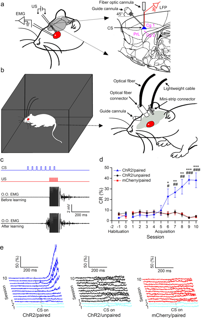
Figure 2. Optogenetic stimulation CS supports the acquisition of associative eyeblink conditioning.

(a) Rats were implanted with stimulating electrodes in the subdermally caudal to the left eye for delivery of unconditioned stimuli (US) and with electrodes for recording the electromyographic (EMG) activity of the ipsilateral orbicularis oculi (O.O.) muscle. An optrode and a guide cannula were targeted to the right caudal mPFC for optical stimulation, for recording local field potentials, and for drug injection. (b) Rats were trained in a sound- and light-attenuating chamber. (c) Upper panel: the conditioning paradigm illustrating the timing of the CS and the US. Middle panel: representative O.O. EMG before learning. Lower panel: representative O.O. EMG after learning. (d, e) the CR% (d) and EMG response topographies (e) across two habituation and ten acquisition training sessions in ChR2/paired, ChR2/unpaired, and mCherry/paired groups ( = 8 rats each; * and # indicate significant differences between the ChR2/paired group and the ChR2/unpaired and mCherry/paired groups; *or #P < 0.05, ** or ##P < 0.01, *** or ###P < 0.001; two-way ANOVA with repeated measures followed by Tukey post-hoc test). Data are represented as mean ± s.e.m. The rat drawing was drawn by Guang-yan Wu according to the present experiment.
