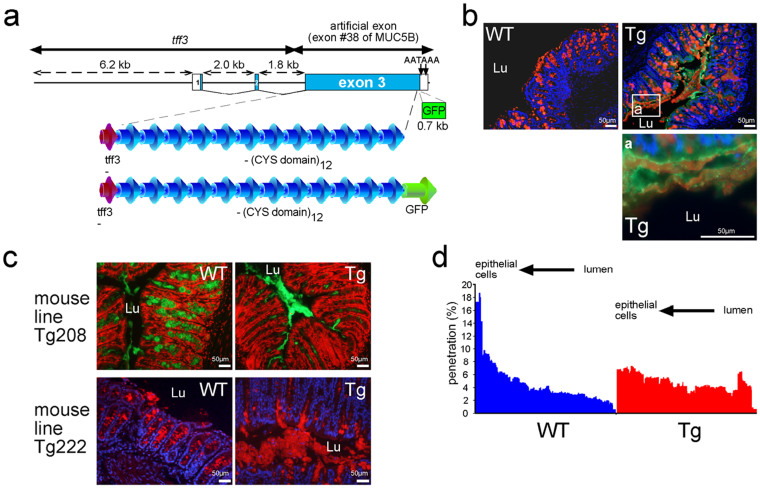
Figure 1. The transgene is secreted into intestinal mucus.
(a). Schematic map of the two DNA fragments that were developed and employed for transgenic (Tg) mice generation and deduced peptide organization. The three exons (exons 1, 2 and 3) are indicated by boxes and numbered. White boxes represent the 5′ and 3′UTR regions. (b). Representative immunohistochemistry of colonic tissue sections of wild-type (WT) and Tg mice. The Tg product is visualized in green and Muc2 is visualized in red using the UEA1 lectin. Cells were counterstained with Hoechst 33258 (blue). (c). Representative IHC of colonic tissue sections of the two Tg mouse lines. Cells were counterstained either with propidium iodide in red (Tg208) or with Hoechst 33258 in blue (Tg222). Muc2 is visualized using an anti-Muc2 antibody in green (Tg208) or in red (Tg222). The colonic mucus was conserved in the two Tg mouse lines. Lu, lumen. (d). The distribution (%) of fluorescent beads loaded at the mucus surface into the colonic mucus in wild-type (WT; n = 12) and Tg (n = 11) mice was studied by confocal microscopy, which showed that the Tg mucus was less penetrable.

