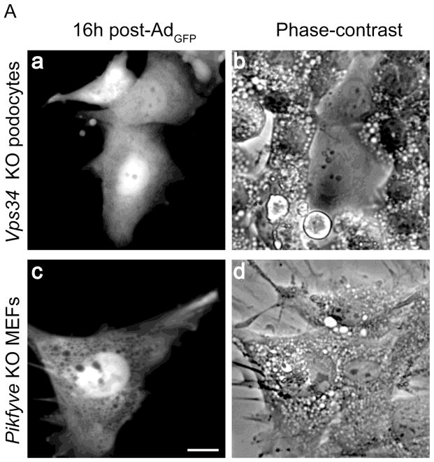
Fig. 5.
Transient activation of class I PI3Ks selectively delays the cytoplasmic vacuolation only in Vps34 KO podocytes. A and B, on day 4 post-Ad-Cre treatment Vps34 KO podocytes and Pikfyve KO MEFs were infected with either AdGFP (A) or AdGFP-PIKfyvewt (B) at MOI=10, as described under “Materials and methods”. 16–36 hours post-infection, cells were observed live by fluorescence and phase-contrast microscopy monitoring the GFP signals and the vacuolation phenotype in both infected [GFP(+)] and noninfected [GFP(−)] cells. A, fluorescence and phase-contrast images of representative cells at 16 h post-infection showing AdGFP -infected Vps34 KO podocytes (panels a and b) that did not exhibit cytoplasmic vacuoles in contrast to the GFP(−) neighboring cells and AdGFP-infected Pikfyve KO MEFs, in which the vacuolation phenotype was unaltered (panels c and d). See C for quantification. Bar, 10 μm. B, fluorescence and phase-contrast images of representative cells at 16 h post-infection with AdGFP-PIKfyvewt showing the absence of vacuolation in AdGFP-PIKfyvewt-expressing Vps34 KO podocytes (panels a and b) and Pikfyve KO MEFs (panels c and d). In both cell lines the aberrant vacuoles persisted in the GFP(−) neighboring cells. See D for quantification. Bar, 10 μm. C and D, Quantification of vacuolation in the non-infected GFP(−) control cells (white bars) and GFP(+)-infected condition (grey bars) as percentage of total GFP(−) control cells and total GFP(+) cells, respectively by counting >100 cells per condition in three separate experiments. *, different vs. GFP(−) cells at the same time point; #, different vs. the 16h time point, P<0.001.


