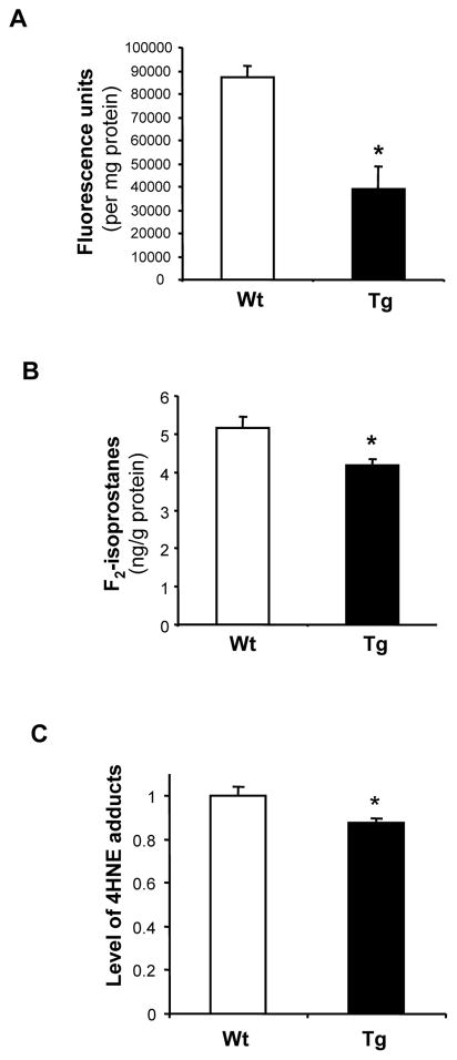
Figure 3. Reduced cellular ROS and oxidative damage in Tg(PRDX3) mice.
A. Levels of cellular ROS in skin fibroblasts from Tg(PRDX3) mice and Wt mice were measured by a DCF fluorescence method. The data are presented as mean ± SEM of data obtained from three independent lines. *: P< 0.05. n=5
B. Levels of F2-isoptostanes in liver tissues of Wt and Tg(PRDX3) were determined as described by Ran et al (2007). The data are expressed as mean ± SEM. *P<0.05. n=5
C. 4-HNE adducts in mitochondria proteins from liver tissues of Tg(PRDX3) mice and Wt mice were determined by Western blots and quantified as described in Methods. The mean value of 4-HNE adducts obtained from Wt mice was artificially assigned as 1, and the relative values are presented as mean ± SEM. *: P<0.05. n= 4–5.

