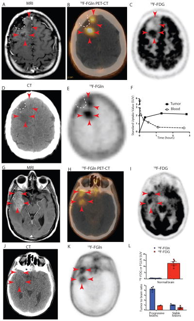
Fig 5. 18F-FGln shows uptake in human gliomas undergoing progression.
A–F. Images from patient #5
A. T1-weighted MRI with contrast enhancement from a 42-year-old IDH1-mutant oligodendroglioma patient showing tumor with minimal gadolinium enhancement (red arrows) along surgical cavity (indicated by white dotted line).
B. Fusion 18F-FGln PET-CT showing 18F-FGln uptake in areas corresponding to tumor (red arrows).
C. 18F-FDG PET image from the same patient showing high background brain avidity and tumor uptake in the posterior part of the tumor (3 red arrows), but not in the anterior portion (2 red arrows).
D. CT scan used to generate the PET-CT fusion image in B.
E. 18F-FGln PET showing high uptake in tumor with minimal uptake in the surrounding brain.
F. Time activity curve indicating standard uptake values (SUV) corresponding to tumor (black squares) and blood (clear circles).
G–K. Images from patient # 6
G. T1-weighted MRI with contrast enhancement from a 57-year-old glioblastoma patient showing tumor with gadolinium enhancement (red arrows).
H. Fusion 18F-FGln PET-CT showing 18F-FGln uptake in areas corresponding to tumor.
I. 18F-FDG PET image from same patient showing high background brain avidity and tumor uptake.
J. CT scan used to generate the PET-CT fusion image in H.
K. 18F-FGln PET showing high uptake in tumor with minimal uptake in the surrounding brain.
L. Comparison of 18F-FGln (blue bars) and 18F-FDG (red bars) illustrates differences in background uptake with both ligands in normal brain (top panel) and tumor to brain ratios from 3 clinically stable glioma patients and 3 glioma patients with clinically progressive disease (bottom panel) (see Tables S1 and S2 for details). For all graphs, data are represented as the means ± s.e.m.

