
Figure 1. The tri-partite synapse.
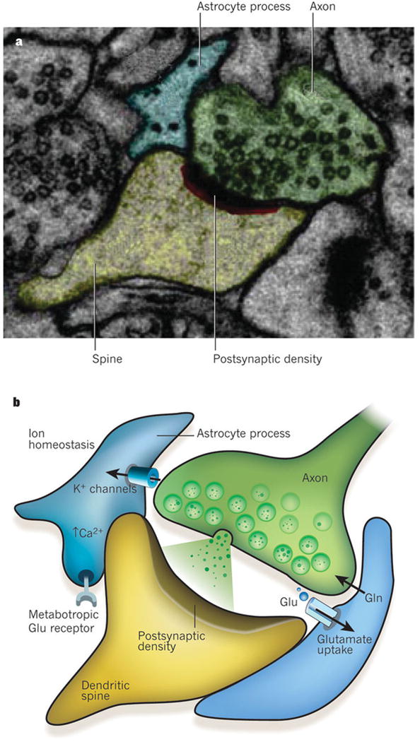
The processes of astrocytes are intimately associated with synapses. This association is both structural and functional. a, Electron micrograph showing a tripartite synapse in the hippocampus. The astrocyte process (blue) ensheaths the perisynaptic area. The axon of the neuron is shown in green, with the dendritic spine in yellow and the postsynaptic density in red and black. Reproduced, with permission, from ref. 22. b, Schematic representation of a tripartite synapse. Perisynaptic astrocyte processes contain transporters that take up glutamate (Glu, green circles) that has been released into the synapse and return it to neurons in the form of glutamine (Gln). Glutamate receptors on astrocytes (such as metabotropic glutamate receptors) sense synaptic glutamate release, which in turn induces a rise in Ca2+ concentration in the astrocytes. One of the main functions of glia at the synapse is to maintain ion homeostasis, for example regulating extracellular K+ concentrations and pH.