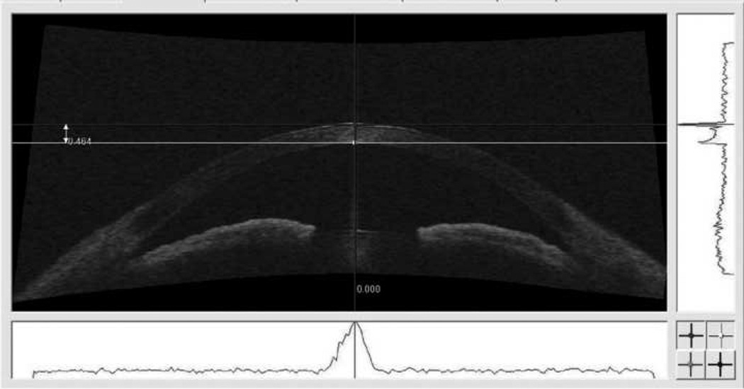
Figure 1.
Cross-sectional image of a subject’s anterior chamber acquired by AS-OCT, demonstrating the method used to measure CCT. The corneal apex was identified after correcting ocular rotation, from the peak of the reflectivity profile on the horizontal axis (below the image). The callipers were then aligned on the peak reflections of the anterior and posterior tissue boundaries of the cornea in the axis of the corneal apex (to the right of the image). The two measurements were averaged for each eye.

