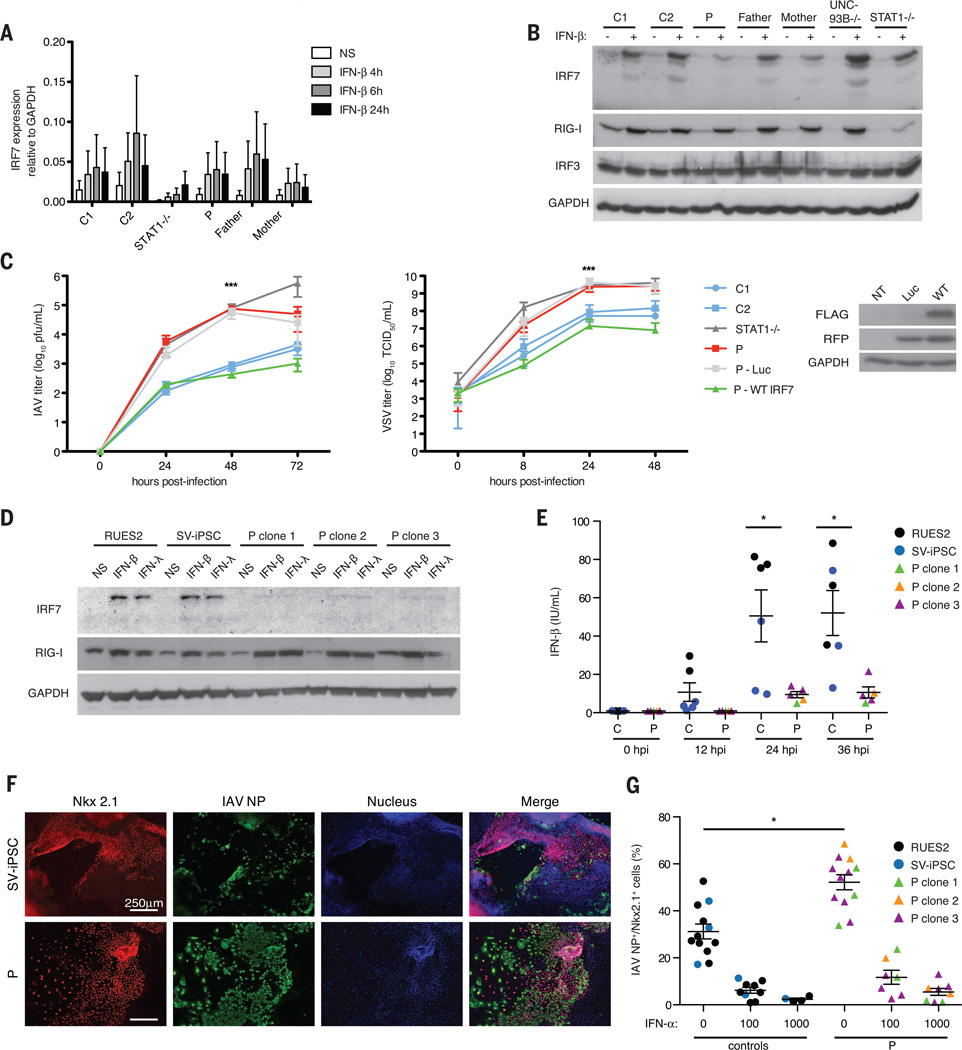
Fig. 4. IRF7-dependent intrinsic immunity is required for control of IAV infection.
(A) IRF7 mRNA induction at indicated time points after IFN-β treatment in F-SV40. Means ± SD of three replicates are shown. (B) IRF7 expression in F-SV40 measured byWestern blot 18 hours after treatment with IFN-β. (C) Virus titers in F-SV40 from P stably transfected luciferase (Luc) or wild-type IRF7 with an internal ribosome entry site–expressed red fluorescent protein, after infection with pH1N1 (MOI = 10) or VSV (MOI = 3). Means ± SD for pH1N1 IAV (n = 3) and VSV (n = 7) are shown. ***P < 0.001 between controls and P as determined by t test. (D) IRF7 expression in IFN-β– or IFN-λ–treated PECs derived from healthy control ESCs (RUES2), SV iPSCs, or three individual clones (clones 1, 2, and 3) of P’s iPSCs as detected by Western blot. (E) IFN-β production in PECs infected with A/PR/8/34-GFP as measured by ELISA at indicated time points. Means ± SD of two independent experiments are shown. *P < 0.05 as determined by t test. (F) Staining of IAV nucleoprotein (NP) (green) and Nkx2.1 (red) in PECs derived from SV-iPSC control and P infected at MOI = 1 with pH1N1 IAV. Cells derived from a single representative clone of P’s iPSCs are shown. (G) Percentage of Nkx2.1+ cells scored as positive for IAV NP, in PECs untreated or treated with IFN-α (100 or 1000 U/ml) for 18 hours and infected with pH1N1 for 24 hours. Means ± SD of two independent experiments are shown. *P < 1 × 10⁻⁶ as determined by χ2 analysis.

