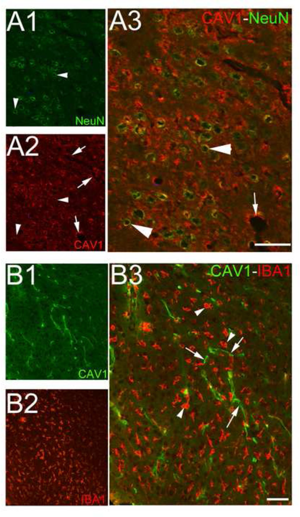
Figure 3. Caveolin 1 expression in neurons but not microglial cells after jTBI.
(A) NeuN labeling (green, A1, A3) and Cav-1 staining (red, A2, A3) co-localized (yellow, arrowheads), suggesting the presence of cav-1 in neurons in the periphery of the lesion. Cav-1 staining (red, A2, A3) is also observed in intracortical microvessels (arrows).
(B) Cav-1 labeling (green, B1, B3, arrows) and IBA1 staining (red, B2, B3, arrowhead), a marker of microglial cells, did not show co-localization.
A, bar = 500µm; B, bar = 100µm

