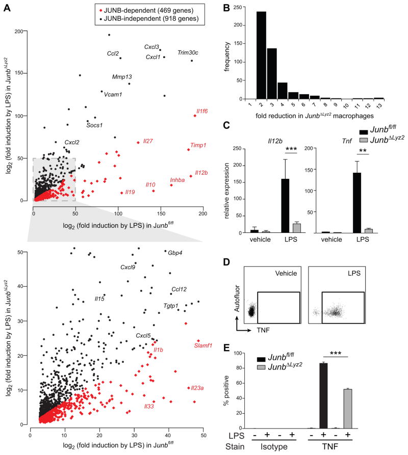
Figure 3. JUNB modulates expression of a cluster of immune-related genes in LPS-treated macrophages.
(A-C) Junbfl/fl or JunbΔLyz2 BMDM were treated for 4 h with LPS. RNA was harvested, amplified, and analyzed by microarray (A, B) or reverse transcribed into cDNA (C). (A) Genes exhibiting < 2-fold difference (black dots) or > 2-fold difference (red dots) in Junbfl/fl versus JunbΔLyz2 macrophages. Results shown are averages from four technical replicates. (B) Histogram of fold-reduction in gene expression for JUNB-dependent genes. (C) Levels of the indicated transcripts were measured by RT-qPCR. Means + SD are shown. Representative results from one of three experiments are shown. **, p < 0.01. ***, p < 0.001 by t-tests. (D) Gating strategy for measurement of intracellular TNF. Isotype controls (not shown) were used as described for pro-IL-1β. (E) Induction of intracellular TNF after 4 h of LPS treatment. Bars represent means + SE for % of BMDM positive for TNF and are representative of three independent experiments.

